Home›Blog ›How do X-ray image intensifiers handle variations in patient anatomy and thickness during fluoroscopic procedures?
How do X-ray image intensifiers handle variations in patient anatomy and thickness during fluoroscopic procedures?
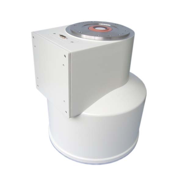
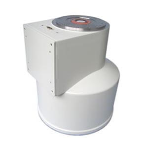
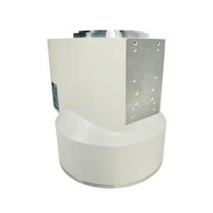
X-ray image intensifiers utilize various techniques to manage variations in patient anatomy and thickness during fluoroscopic procedures. Automatic exposure control (AEC) systems adjust X-ray output based on the thickness of the anatomy being imaged, ensuring optimal image quality while minimizing unnecessary radiation exposure. Additionally, dynamic range compression (DRC) techniques adjust image contrast to accommodate variations in tissue density, allowing for clear visualization of structures across different anatomical regions. Beam filtration systems may also be employed to selectively attenuate low-energy X-rays, reducing radiation dose to the patient while maintaining image quality. Furthermore, real-time feedback mechanisms continuously monitor image quality and radiation dose, enabling adjustments during the procedure to account for changes in patient positioning or anatomy. These technologies work in tandem to optimize image quality while minimizing radiation exposure, ensuring safe and effective fluoroscopic procedures across a range of patient anatomies and thicknesses. We are a manufacturer of X-ray machines and their accessories. If you have any needs for image intensifiers, please feel free to contact us. Whatsapp:+86 18953679166. Email: service@newheek.com
Author:Image Intensifier
Product Category
News
- Image intensifier: the “magnifying glass” of medical images
- X-ray image intensifier television system
- Beyond the Basics: Understanding High-Voltage Cables for X-Ray Machines (75KV & 90KV)
- Elbow 75KV High Voltage Cable: The Hidden Backbone of Reliable X-Ray Machine Performance
- NK-15XZ-Ⅱ 6-inch X-Ray Image Intensifier: The Quiet Workhorse of Real-Time Imaging
Contact us
Tel: (+86) 18953679166
Whatsapp: +86 18953679166
Email: service@newheek.com
Company: Weifang Newheek Electronic Technology Co., Ltd.
ADD: E Building of Future Star Scientific Innovation Industrial Zone of No.957 Wolong East Street, Yulong Community, Xincheng Sub-District Office, Weifang Hi-tech Zone, Shandong Province, China