Home›Blog ›Are there any limitations to using an X-ray image intensifier in terms of imaging depth?
Are there any limitations to using an X-ray image intensifier in terms of imaging depth?
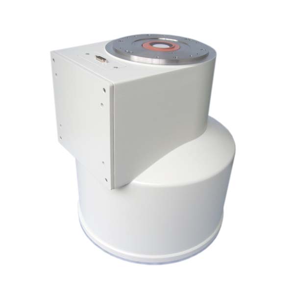
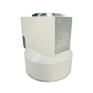
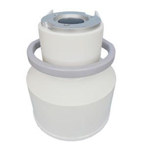
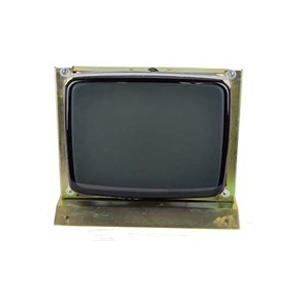
An X-ray image intensifier is a device commonly used in medical imaging to visualize X-ray images in real-time. While it offers advantages such as increased image brightness and the ability to provide dynamic imaging, there are limitations to its use, particularly in terms of imaging depth. Here are some considerations:
1. Limited Depth Penetration:
– X-ray image intensifiers are generally more suitable for imaging structures near the surface of the body. They are commonly used in procedures such as fluoroscopy, which involves real-time imaging of moving structures like the gastrointestinal tract or blood vessels.
2. Decreased Image Quality with Depth:
– As X-rays pass through the body, they are attenuated (absorbed) by the tissues. Deeper structures may receive fewer X-rays, resulting in a decrease in image quality. This is known as the “penumbra effect.” It can lead to reduced visibility of fine details in deeper tissues.
3. Limited for Deep Organs:
– When imaging deep-seated organs or structures, such as those within the chest or abdomen, the X-ray image intensifier may not provide optimal image quality. This is particularly true for structures that are located far from the X-ray source.
4. Resolution and Magnification Challenges:
– While image intensifiers can provide enhanced brightness and magnification, there are limits to their spatial resolution. Fine details in deep structures may be challenging to visualize with the same clarity as those near the surface.
5. Alternative Imaging Modalities:
– For imaging structures at greater depths, other modalities like computed tomography (CT) or magnetic resonance imaging (MRI) may be more suitable. These modalities offer cross-sectional imaging and can provide detailed information about internal structures regardless of their depth.
It’s essential to consider the specific clinical scenario and imaging requirements when choosing the appropriate imaging modality. Radiologists and medical professionals carefully select the imaging technique that balances the need for depth penetration, resolution, and real-time imaging based on the clinical question and the anatomy of interest. Additionally, advancements in technology may lead to improvements in imaging depth capabilities in certain situations. Whatsapp:+86 18953679166. Email: service@newheek.com
Author:Image Intensifier
Product Category
News
- Image intensifier: the “magnifying glass” of medical images
- X-ray image intensifier television system
- Beyond the Basics: Understanding High-Voltage Cables for X-Ray Machines (75KV & 90KV)
- Elbow 75KV High Voltage Cable: The Hidden Backbone of Reliable X-Ray Machine Performance
- NK-15XZ-Ⅱ 6-inch X-Ray Image Intensifier: The Quiet Workhorse of Real-Time Imaging
Contact us
Tel: (+86) 18953679166
Whatsapp: +86 18953679166
Email: service@newheek.com
Company: Weifang Newheek Electronic Technology Co., Ltd.
ADD: E Building of Future Star Scientific Innovation Industrial Zone of No.957 Wolong East Street, Yulong Community, Xincheng Sub-District Office, Weifang Hi-tech Zone, Shandong Province, China