Home›Blog ›Can an X-ray image intensifier be used for fluoroscopy imaging?
Can an X-ray image intensifier be used for fluoroscopy imaging?
Yes, an X-ray image intensifier is commonly used in fluoroscopy imaging. Fluoroscopy is a medical imaging technique that provides real-time, dynamic imaging of the internal structures of a patient. It involves the use of a continuous X-ray beam to visualize moving structures, such as the digestive system, blood vessels, and joints.
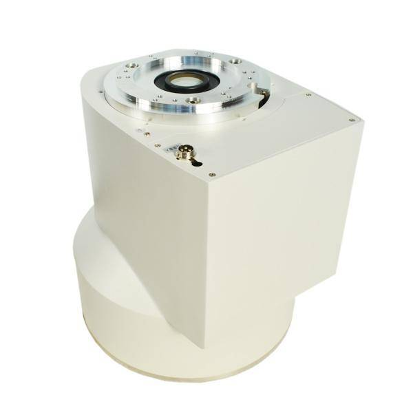
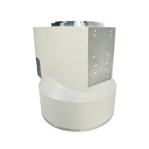
An X-ray image intensifier is a key component in fluoroscopy systems. It is designed to amplify the X-ray signal and convert it into a visible image. The basic components of an X-ray image intensifier include:
1. Input Phosphor:
– The X-ray beam first interacts with an input phosphor, which absorbs the X-rays and emits light in response.
2. Photocathode:
– The emitted light then strikes a photocathode, a photoemissive material that converts the light into electrons.
3. Electrostatic Focusing:
– These electrons are accelerated and focused by an electrostatic field toward a small output phosphor.
4. Output Phosphor:
– The accelerated electrons strike the output phosphor, producing a brighter and magnified image that is visible to the human eye or can be captured by a camera.
5. Fluoroscopic Display:
– The resulting image is displayed in real-time on a monitor, allowing the continuous observation of anatomical structures and the movement of contrast agents.
The use of an X-ray image intensifier in fluoroscopy provides several advantages, including:
– Real-time Imaging: Fluoroscopy allows dynamic imaging in real-time, enabling physicians to visualize the movement and function of internal structures during medical procedures.
– Enhanced Sensitivity: The image intensifier significantly amplifies the X-ray signal, improving the visibility of low-intensity structures and reducing the radiation dose required for imaging.
– High Resolution: The magnification capabilities of the image intensifier contribute to higher spatial resolution in fluoroscopic images.
– Versatility: Fluoroscopy systems with image intensifiers are versatile and can be used in various medical procedures, such as gastrointestinal examinations, cardiac catheterizations, and interventional radiology procedures.
It’s important to note that while X-ray image intensifiers are widely used, newer technologies such as flat-panel detectors are also being employed in fluoroscopy systems. These detectors offer advantages such as improved image quality, lower radiation dose, and a flat, compact design. However, X-ray image intensifiers continue to be used in many fluoroscopy systems, especially in existing installations. Whatsapp:+86 18953679166. Email: service@newheek.com
Author:Image Intensifier


Product Category
News
- Image intensifier: the “magnifying glass” of medical images
- X-ray image intensifier television system
- Beyond the Basics: Understanding High-Voltage Cables for X-Ray Machines (75KV & 90KV)
- Elbow 75KV High Voltage Cable: The Hidden Backbone of Reliable X-Ray Machine Performance
- NK-15XZ-Ⅱ 6-inch X-Ray Image Intensifier: The Quiet Workhorse of Real-Time Imaging
Contact us
Tel: (+86) 18953679166
Whatsapp: +86 18953679166
Email: service@newheek.com
Company: Weifang Newheek Electronic Technology Co., Ltd.
ADD: E Building of Future Star Scientific Innovation Industrial Zone of No.957 Wolong East Street, Yulong Community, Xincheng Sub-District Office, Weifang Hi-tech Zone, Shandong Province, China