Home›Blog ›Application of image intensifier in fluoroscopy
Application of image intensifier in fluoroscopy
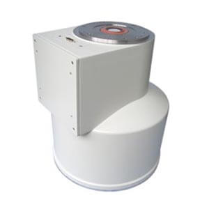
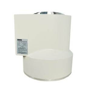
After image intensifier in fluoroscopy conversion X-ray photoelectron is accelerated by high pressure. It is formed on the output plane by electron lens composed of cluster electrode and anode.
A cylindrical crystal with optical fiber structure is formed on the input plane, which is used to suppress the optical diffusion and improve the spatial frequency characteristics. The output plane forms a fluorescent film directly through the output plane. In addition, high contrast images can be obtained by using the antireflective layer.
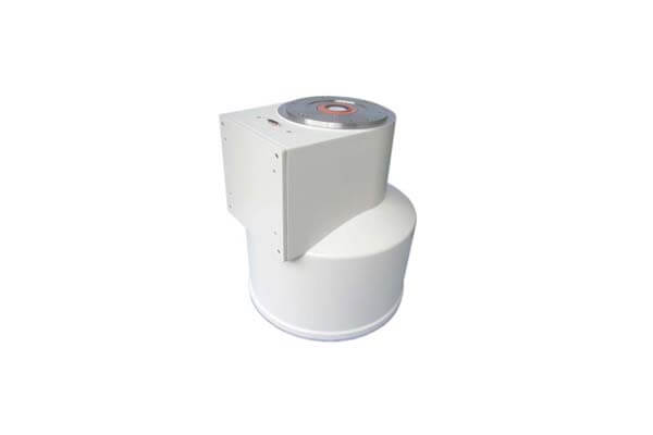
The image intensifier USES X-ray as the light source, and the object taken by the camera is the fluorescent image on the intensifier, which is different from other TV systems that use visible light as the light source.
As for your application, image intensifier in fluoroscopy liberated your intensifier from the dark room.Before doctors must wear red glasses after about five minutes of dark adaptation, in total darkness of the indoor observation screen image.The image is clear, the contrast is good, is advantageous to discover the lesion, enhanced the work efficiency and the diagnosis rate.
Newheek image intensifier in fluoroscopy can replace Toshiba, thales, philips and more.
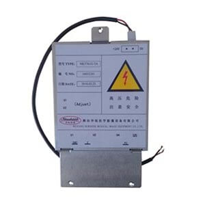
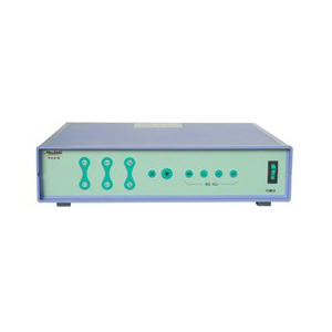
Newheek can also provide quality testing for your image intensifier in fluoroscopy.
Author:Glinda
Product Category
News
- Beyond the Basics: Understanding High-Voltage Cables for X-Ray Machines (75KV & 90KV)
- Elbow 75KV High Voltage Cable: The Hidden Backbone of Reliable X-Ray Machine Performance
- NK-15XZ-Ⅱ 6-inch X-Ray Image Intensifier: The Quiet Workhorse of Real-Time Imaging
- Why Are X-ray Image Intensifiers Falling Behind?
- Real-World Case Study: NK5761-TA Power Supply Replacement for Thales TH9464 6” Image Intensifier
Contact us
Tel: (+86) 18953679166
Whatsapp: +86 18953679166
Email: service@newheek.com
Company: Weifang Newheek Electronic Technology Co., Ltd.
ADD: E Building of Future Star Scientific Innovation Industrial Zone of No.957 Wolong East Street, Yulong Community, Xincheng Sub-District Office, Weifang Hi-tech Zone, Shandong Province, China