Home›Blog ›Image intensifier: the “magnifying glass” of medical images
Image intensifier: the “magnifying glass” of medical images
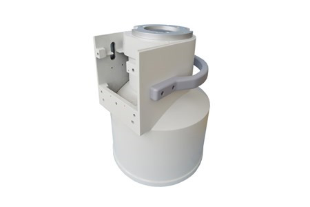
The image intensifier can be called the “magnifying glass” of medical images. It can amplify weak X-ray signals ten thousand times, convert blurry perspective images into high-definition images that are clearly visible in a bright room, and significantly reduce radiation dose. It is an indispensable core imaging component in interventional surgeries and routine radiographs.
1. Image intensifier: From “supporting role” to “Mr. Key”
Image intensifiers are often misunderstood as “auxiliary equipment,” but their role goes far beyond their “supporting role.” Just like mobile phone cameras require the cooperation of optical image stabilization and night scene mode algorithms, medical imaging equipment (such as X-ray machines and CT) also require image intensifiers to improve imaging quality. Its core function is to convert weak X-ray signals into visible light, and then enhance image details through electronic amplification technology, allowing doctors to see clearly 0.1 mm-level blood vessels or bone microcracks. This technology has greatly improved the accuracy of early lung cancer screening, orthopedic surgery navigation and other scenarios, becoming a “quality amplifier” for modern medical imaging.
2. The “golden partner” relationship with the main device
The relationship between the image intensifier and the main device is more like “coffee and a companion” – it has limited functions when it exists alone, but can release ideal effects when combined. For example: In digital subtraction angiography (DSA), the main device is responsible for emitting X-rays, and the image intensifier is responsible for capturing dynamic images of blood flow in real time, and using algorithms to eliminate interference from bones and muscles to generate clear three-dimensional maps of blood vessels. This synergy shortens the time of cardiac interventional surgery from 2 hours to 40 minutes and reduces the patient’s radiation exposure by 60%. In pediatric applications, image intensifiers can reduce the imaging dose of children’s bones to 1/3 of adults while maintaining image clarity, making it a “child-friendly” medical technology.
3. Beyond Assistance: Independent Value and Scenario Expansion
The value of image intensifiers has exceeded traditional auxiliary positioning. In mobile medical scenarios, it is integrated into portable X-ray machines, allowing grassroots hospitals to complete inspections that could only be done in tertiary hospitals in the past; in the field of industrial non-destructive testing, it is used to detect internal cracks in aircraft blades with an accuracy of 0.05 mm; and even in the security field, it can enhance the image details of nighttime surveillance and help police quickly identify suspects. These applications prove that the image intensifier is not only an “auxiliary tool”, but also a core technology for cross-domain image processing. Its value depends on how it is combined with specific scenes – just like each blade of a Swiss army knife, it is an accessory alone, but when combined, it is a sharp tool to solve problems.
Author:Image Intensifier
Product Category
News
- Medical CCD Camera | Reliable Component for C-Arm System Refurbishment
- C arm image intensifier | 6/9/12 inch medical X-ray image intensifier replacement parts
- Replacement High Voltage Power Supply for X-Ray Image Intensifier: A Complete Buyer’s Guide
- Cost-Effective Medical Image Intensifier: High-Resolution Low-Distortion for Budget-Conscious Facilities
- Image Intensifier: A Practical Core Device for X-Ray Imaging
Contact us
Tel: +86 19015366638
Whatsapp: +86 19015366638
Email: newheekcn@163.com
Company: Weifang Newheek Electronic Technology Co., Ltd.
ADD: E Building of Future Star Scientific Innovation Industrial Zone of No.957 Wolong East Street, Yulong Community, Xincheng Sub-District Office, Weifang Hi-tech Zone, Shandong Province, China