Home›Blog ›The structure and imaging process of Xray image intensifier
The structure and imaging process of Xray image intensifier
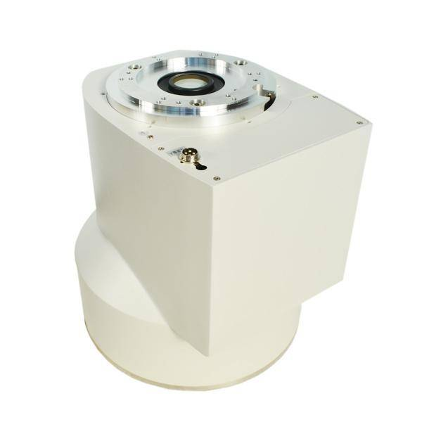
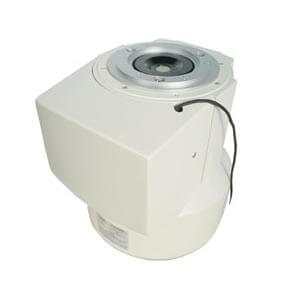
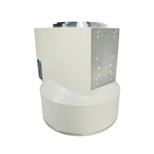
Xray image intensifiers are usually cylindrical, with internal vacuum vessels containing multiple components.
X-ray sensitive input fluorescent screen converts invisible X-ray photon images into visible light images.
A photon-excited photocathode emits an electronic image.
Electrons are accelerated by thousands of volts and focused on a fluorescent screen to create a visible image.
The visible light image reflects the details of the latent image, and the brightness is greatly enhanced.
The image can be observed by a television camera system with high sensitivity or sent to a computer for processing and recognition by a CCD camera and other digital acquisition systems.
The Xray image intensifier is mainly composed of the following parts: input screen, input scintillation crystal, photocathode, vacuum tube and focusing electrode, output scintillation body and output screen.
Newheek specializes in the production of X-ray machines and radiology equipment, the main products are: portable X-ray machines, digital X-ray machines, X-ray collimators, Xray image intensifiers, etc. If your company is in need of such equipment, welcome to send email and get in touch with us!
Author:肖恩
Product Category
News
- Beyond the Basics: Understanding High-Voltage Cables for X-Ray Machines (75KV & 90KV)
- Elbow 75KV High Voltage Cable: The Hidden Backbone of Reliable X-Ray Machine Performance
- NK-15XZ-Ⅱ 6-inch X-Ray Image Intensifier: The Quiet Workhorse of Real-Time Imaging
- Why Are X-ray Image Intensifiers Falling Behind?
- Real-World Case Study: NK5761-TA Power Supply Replacement for Thales TH9464 6” Image Intensifier
Contact us
Tel: (+86) 19015366638
Whatsapp: +86 19015366638
Email: newheekcn@163.com
Company: Weifang Newheek Electronic Technology Co., Ltd.
ADD: E Building of Future Star Scientific Innovation Industrial Zone of No.957 Wolong East Street, Yulong Community, Xincheng Sub-District Office, Weifang Hi-tech Zone, Shandong Province, China