Home›Blog ›X-ray machine fluoroscopy image intensifier size
X-ray machine fluoroscopy image intensifier size
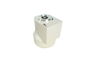
The working principle of the image intensifier: the transmitted X-ray (X-ray image) passing through the human body is irradiated on the input screen of the image intensifier to obtain a fluorescent image with weaker brightness, and then a size is obtained on the output screen after being enhanced by the image intensifier A reduced fluorescent image with a brightness that is ten million times stronger than that on the input screen. After the fluorescent image on the output screen is transmitted and corrected by the optical system, it is picked up by the camera tube. The video current signal output from the camera tube is amplified by the preamplifier, and the controller performs image signal control, processing and amplification to obtain the full TV signal. Go to the monitor and display the X-ray fluoroscopy image on the monitor phosphor screen.
Image intensifier structure: It is composed of an image multiplier tube, a tube container, a dedicated high-voltage power supply built in the tube container, and a low-voltage power supply for driving the high-voltage power supply.
Author:Lillian


Product Category
News
- Beyond the Basics: Understanding High-Voltage Cables for X-Ray Machines (75KV & 90KV)
- Elbow 75KV High Voltage Cable: The Hidden Backbone of Reliable X-Ray Machine Performance
- NK-15XZ-Ⅱ 6-inch X-Ray Image Intensifier: The Quiet Workhorse of Real-Time Imaging
- Why Are X-ray Image Intensifiers Falling Behind?
- Real-World Case Study: NK5761-TA Power Supply Replacement for Thales TH9464 6” Image Intensifier
Contact us
Tel: (+86) 18953679166
Whatsapp: +86 18953679166
Email: service@newheek.com
Company: Weifang Newheek Electronic Technology Co., Ltd.
ADD: E Building of Future Star Scientific Innovation Industrial Zone of No.957 Wolong East Street, Yulong Community, Xincheng Sub-District Office, Weifang Hi-tech Zone, Shandong Province, China