Home›Blog ›The image intensifier on the C-arm X-ray machine is used for replacement
The image intensifier on the C-arm X-ray machine is used for replacement
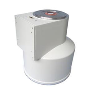
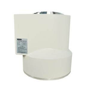
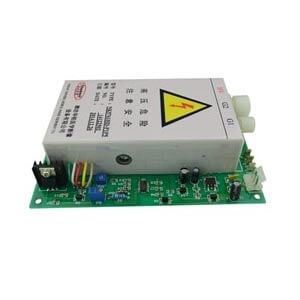
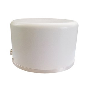
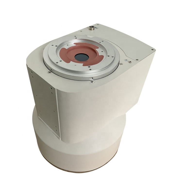
I recently received a customer’s inquiry about replacing the image intensifier on the c-arm X-ray machine. The customer wanted a 6-inch image intensifier and asked if we had it in stock. After checking the inventory, we would reply to the customer that we have it in stock. Image intensifier structure: It is composed of an intensifier tube, a tube container, a power supply, an optical system, and a support (support) part.
The main part of the image intensifier is the image intensifier tube, which has an input screen (receiving X-ray radiation to generate electron flow) and an output screen (receiving electron bombardment to emit light), which enables the former to enhance images with thousands of times the brightness of the image on the output screen.
The resolution of the image intensifier is limited by the resolution of the input and output phosphor screen and the ability of the focusing electrode to maintain the image when the image is transferred from the input screen to the output screen. The patient’s motion and the limited size of the focal spot cause image loss of sharpness. In addition, the quality of the fluoroscopy image is also affected by the statistical fluctuation of the number of X-rays hitting the input screen. The resolution, brightness, and contrast of the image produced by the image intensifier are the largest at the center of the image, and gradually decrease toward the periphery. The brightness reduction of the image along the peripheral direction is usually no more than 25%.
The above is all about the impact enhancer, welcome to consult.
Author:Lillian
Product Category
News
- A customer in Y* asked for an image intensifier
- An inquiry about image intensifier
- A medical equipment maintenance company in Jiangsu consulted Newheek for the image intensifier
- Z* customers inquire about image intensifier maintenance of Newheek
- Lebanese customers inquire about Newheek for image intensifiers
Contact us
Tel: (+86) 18953679166
Whatsapp: +86 18953679166
Email: service@newheek.com
Company: Weifang Newheek Electronic Technology Co., Ltd.
ADD: E Building of Future Star Scientific Innovation Industrial Zone of No.957 Wolong East Street, Yulong Community, Xincheng Sub-District Office, Weifang Hi-tech Zone, Shandong Province, China
