Home›Blog ›Image intensifier development process
Image intensifier development process
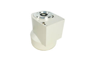
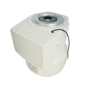
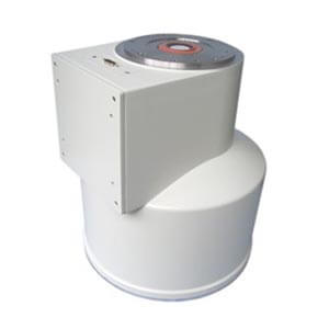
Do you know the history of image intensifier TV system? Simply talk about the history of the image intensifier TV system. In the early fluoroscopy technique, the patient’s X-ray directly hit the screen. The visible light emitted by each region of the screen is related to the rate of energy deposited by the incident X-ray. The radiologist observes the visible light image on the screen at a distance of 10 or 15 inches. There is a thin lead glass plate behind the screen to protect the radiologist from the X-rays that pass through the screen.

In the 1940s, radiologists realized that the low visibility of images in fluoroscopy was associated with dim images in early fluoroscopy. They emphasize the need for brighter fluorescent images and encourage the development of image intensifiers. The image intensifier increases the brightness of the fluorescent image, and observers can use clear vision (cone cells) instead of the scotopic (rod cells) required in earlier fluoroscopy. Fluorescence with image intensifier does not require dark adaptation due to the brighter image. Although image intensifier increases the cost and complexity of fluoroscopy systems, the use of non-image intensifier fluoroscopy is outdated.
Author:Alina
Product Category
News
- A customer in Y* asked for an image intensifier
- An inquiry about image intensifier
- A medical equipment maintenance company in Jiangsu consulted Newheek for the image intensifier
- Z* customers inquire about image intensifier maintenance of Newheek
- Lebanese customers inquire about Newheek for image intensifiers
Contact us
Tel: (+86) 18953679166
Whatsapp: +86 18953679166
Email: service@newheek.com
Company: Weifang Newheek Electronic Technology Co., Ltd.
ADD: E Building of Future Star Scientific Innovation Industrial Zone of No.957 Wolong East Street, Yulong Community, Xincheng Sub-District Office, Weifang Hi-tech Zone, Shandong Province, China


