Home›Blog ›Is Your C-Arm Image Intensifier Actually Failing? 6 Symptoms That Point to Replacement (and 3 That Don’t)
Is Your C-Arm Image Intensifier Actually Failing? 6 Symptoms That Point to Replacement (and 3 That Don’t)
A C-Arm Image Intensifier rarely dies with a dramatic pop. More often, it fades—slowly enough that the clinical team adapts without realizing the system has drifted from its baseline. That gradual decline is exactly what makes the replacement decision difficult. A dark image, a soft edge, or a new artifact could mean the C-Arm Image Intensifier is finished. Or it could mean the generator, the camera chain, or a calibration setting is the real culprit.
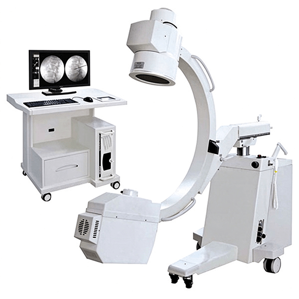
Here is how to tell the difference before you authorize a five-figure part.
How the C-Arm Image Intensifier ages. X-rays strike the input phosphor and release light photons, which liberate electrons from the photocathode. Electron optics accelerate and focus those electrons onto a small output phosphor, producing a bright image for the camera to capture. Every stage degrades at its own rate: the input phosphor loses conversion efficiency, the electron optics drift out of focus, and the output phosphor develops permanent wear from repeated collimated fields. Because the decline is distributed across the tube, the symptoms tend to cluster—and that clustering is what distinguishes a genuine C-Arm Image Intensifier failure from an upstream fault.
Six symptoms that point to replacement
1. You keep increasing technique to hold the same brightness. If the system needs progressively higher kVp or mA to produce an image that used to come easily, the input phosphor is likely losing reactivity. This is the earliest and most reliable sign. It is also the easiest to miss because the generator compensates automatically—until it cannot.
2. Fixed dark spots appear and do not move. Sharp, non-moving dark spots on the image usually mean physical debris or phosphor flaking inside the C-Arm Image Intensifier. Unlike digital artifacts, these do not disappear after a cable reseat or a reboot. If the spots are visible across every field-of-view mode and every C-arm position, the tube itself is the source.
3. Contrast collapses and tissue densities blend together. When a failing vacuum or a degraded output phosphor flattens the image, the fine separation between soft tissue and bone disappears. The image looks washed out rather than simply dim. No amount of window/level adjustment restores the missing contrast because the information is no longer being generated inside the tube.
4. Edge distortion becomes clinically noticeable. Pincushion distortion is inherent to electron-optically coupled tubes, but it should remain stable and minor—typically within the range the system’s correction handles. When the distortion visibly worsens, or when the edges darken unevenly (vignetting), the electron optics or the output window are degrading beyond what calibration can compensate.
5. Localized burn-in or ghosting persists across cases. Static anatomy or tightly collimated fields leave a latent image on the output phosphor that no longer fades between patients. If the same outline follows you from case to case, the output screen has suffered permanent wear. This is irreversible and directly compromises interpretation in subsequent procedures.
6. The symptom changes with magnification mode—and only with magnification mode. A fault that appears or intensifies when switching from 9-inch to 6-inch mode points toward the C-Arm Image Intensifier’s internal electron-optics geometry, not the generator or camera. This is not absolute proof on its own, but combined with any of the symptoms above, it strongly narrows the fault to the tube.
Three symptoms that do not point to replacement
A uniformly dark image that responds to generator output changes. The C-Arm Image Intensifier may be innocent. A failing high-voltage supply, a drifting generator calibration, or a camera gain issue can all darken the image without the tube being at fault. Before condemning the C-Arm Image Intensifier, confirm that X-rays are actually being produced at the expected output.
Artifacts that move or disappear when cables are reseated. Digital artifacts from poor connections mimic tube faults. If the artifact changes with cable manipulation, or vanishes after a system reset, the problem sits in the signal path—not inside the vacuum tube.
Blur or distortion that corrects with focus or collimator adjustment. A dirty mirror, a misadjusted collimating lens, or an out-of-focus optical coupling can produce fuzzy images that look like tube degradation. These are serviceable faults. Rule them out before ordering a replacement.
The practical takeaway
The fastest way to avoid an unnecessary replacement is a simple two-step isolation: confirm X-ray production in front of the C-Arm Image Intensifier, then determine whether the fault persists across every field-of-view mode and C-arm position. If the fault is mode-dependent and accompanied by brightness drift, fixed spots, or persistent ghosting, the C-Arm Image Intensifier is the likely source. If the fault is mode-independent and responds to generator, cable, or calibration changes, keep looking upstream.
A replacement C-Arm Image Intensifier restores the system only when the C-Arm Image Intensifier is the actual failed component. Document the baseline, isolate the chain, and let the symptom pattern—not the first dark image—drive the decision.Request a Compatibility Check
Author:Image Intensifier
Product Category
News
- 9-Inch vs. 12-Inch Image Intensifier: Why Tube Size Alone Is the Wrong Way to Choose a Replacement
- Is Your C-Arm Image Intensifier Actually Failing? 6 Symptoms That Point to Replacement (and 3 That Don’t)
- output phosphor of image intensifier
- Image Intensifier Market
- Compatible X-ray II High Tension Power Unit
Contact us
Tel: +86 19015366638
Whatsapp: +86 19015366638
Email: newheekcn@163.com
Company: Weifang Newheek Electronic Technology Co., Ltd.
ADD: E Building of Future Star Scientific Innovation Industrial Zone of No.957 Wolong East Street, Yulong Community, Xincheng Sub-District Office, Weifang Hi-tech Zone, Shandong Province, China