Home›Blog ›The changing process of image intensifier function structure
The changing process of image intensifier function structure
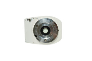
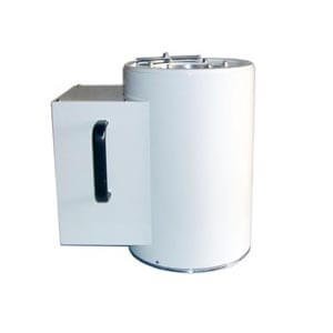
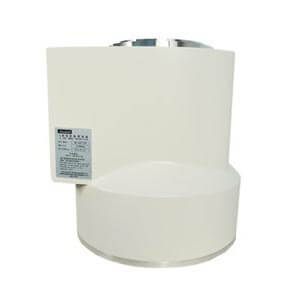
The image intensifiers currently used in most hospitals are the first generation of image conversion equipment for bright room fluoroscopy, X-ray dose reduction and compartment operations. From the appearance, the image intensifier is a large glass tube with a black dressing on the surface as a light sealing layer, and a high vacuum is maintained in the tube.
There is an input screen with a large area at the front end of the tube, and a layer of phosphor is coated on the input screen. The thicker the phosphor layer, the stronger the brightness, but this will reduce the resolution due to light scattering and reflection; the thinner the phosphor layer, the higher the resolution, but the brightness is reduced. In order to solve this contradiction, in recent years, new image intensifiers have adopted a relatively high atomic number cesium iodide phosphor input screen. Compared with the zinc cadmium sulfide phosphors used earlier, cesium iodide phosphors have the advantages of high X-ray absorptivity, high fluorescence efficiency, high image resolution and good matching with the photocathode spectrum. Like ordinary fluoroscopic screens, this screen absorbs X-ray photons with image information to produce visible fluorescent images.
Close to the input screen is the photocathode (there is a thin transparent layer between the two); the antimony-cesium type photocathode, when the photon of the phosphor image of the input screen irradiates the photocathode surface, the other side emits photoelectrons to form Electronic image, complete the photo-electric conversion process.

Author:Lillian
Product Category
News
- A customer in Y* asked for an image intensifier
- An inquiry about image intensifier
- A medical equipment maintenance company in Jiangsu consulted Newheek for the image intensifier
- Z* customers inquire about image intensifier maintenance of Newheek
- Lebanese customers inquire about Newheek for image intensifiers
Contact us
Tel: (+86) 18953679166
Whatsapp: +86 18953679166
Email: service@newheek.com
Company: Weifang Newheek Electronic Technology Co., Ltd.
ADD: E Building of Future Star Scientific Innovation Industrial Zone of No.957 Wolong East Street, Yulong Community, Xincheng Sub-District Office, Weifang Hi-tech Zone, Shandong Province, China


